Teaching in courses:
Research Group
CFU

Group leader: Jørgen Arendt Jensen
Center for Fast Ultrasound Imaging (CFU)
The Center for Fast Ultrasound Imaging (CFU) was inaugurated in 1998 by Professor Jørgen Arendt Jensen and has pioneered a range of innovations in medical ultrasound. The major contributions include ultrasound simulation with development and maintenance of the gold standard Field II program, and development of ultrasound research scanners including the pioneering RASMUS and SARUS systems.
CFU also invented transverse oscillation vector flow imaging (VFI), which was commercially introduced on BK Medical scanners for the worlds first clinical VFI. Major results have also been presented within anatomic and vector flow synthetic aperture imaging in 2-D with commercial arrays and in 3-D with row-column arrays, which has resulted in 27 sold patents.
CFU is currently sponsored by an 10 million Euro ERC Synergy grant for super resolution imaging and houses one professor, a senior researcher, 2 postdocs and 15 technical and clinical PhD students. We have collaborations with several departments at DTU as well as the University of Copenhagen and University hospitals in the Copenhagen region.
We have extensive research facilities in terms of 5 research scanners (SARUS and 4 Verasonics scanners), robot measurement systems, flow rigs, extensive collection of probes and commercial scanners and several cluster and GPU computers for finite element and ultrasound simulation and image processing. The USB section also houses the MEMS group, with full clean-room facilities for fabricating and testing 1-D and 2-D CMUT probes, and the Biomechanics group for advanced finite element flow simulations.
News from CFU
Show all newsPhDs and Postdocs
Discover all of our PhD projects here
Research
Research at CFU spans the development of algorithms and their implementation, development of equipment, pre-clinical trials and efficacy evaluation of the new techniques. Several major results have been obtained, published and patented.
To read more about the individual research areas, choose in the menu below.
Ongoing research projects include:
- Synthetic Aperture Compound Imaging 3D Vector Flow Imaging
- 3D Vector Flow Imaging
- Synthetic aperture flow imaging using a dual beam former approach
- Non-linear SA Imaging
- Micromachined integrated transducers for ultrasound imaging
One of our successes is a research scanner for SA imaging - RASMUS. The scanner can send out advanced coded fields and acquire data from all transducer channels in real time for clinical imaging. The system generates 5 GBytes of data per second, and this can be stored in the internal 16 GBytes memory for the system. This makes it possible to make clinical, real-time data acquisition and storing the data in a 32 processor Linux cluster for processing.
All developed methods have been investigated on data from human volunteers using this system, and the methods have been shown to function for clinical imaging.
A simulation package, Field II, for ultrasound imaging has also been developed at CFU and distributed on the web. The software is considered a standard for simulating ultrasound systems and is used by numerous research groups and market leading companies (Philips, Siemens, General Electric, Esaote, B-K Medical, Aloka, Vermon).
Engineers and researchers from the private industry are active collaborators suggesting specific topics for M.Sc. or Ph.D. projects and contributing with their experience and expertise in applied research and development as well as acting as external supervisors.
Research Areas
Flow estimation
Ultrasonic flow estimation is widely used to evaluate the blood flow in the human body. This allows medical doctors to evaluate the function of the cardiovascular system.

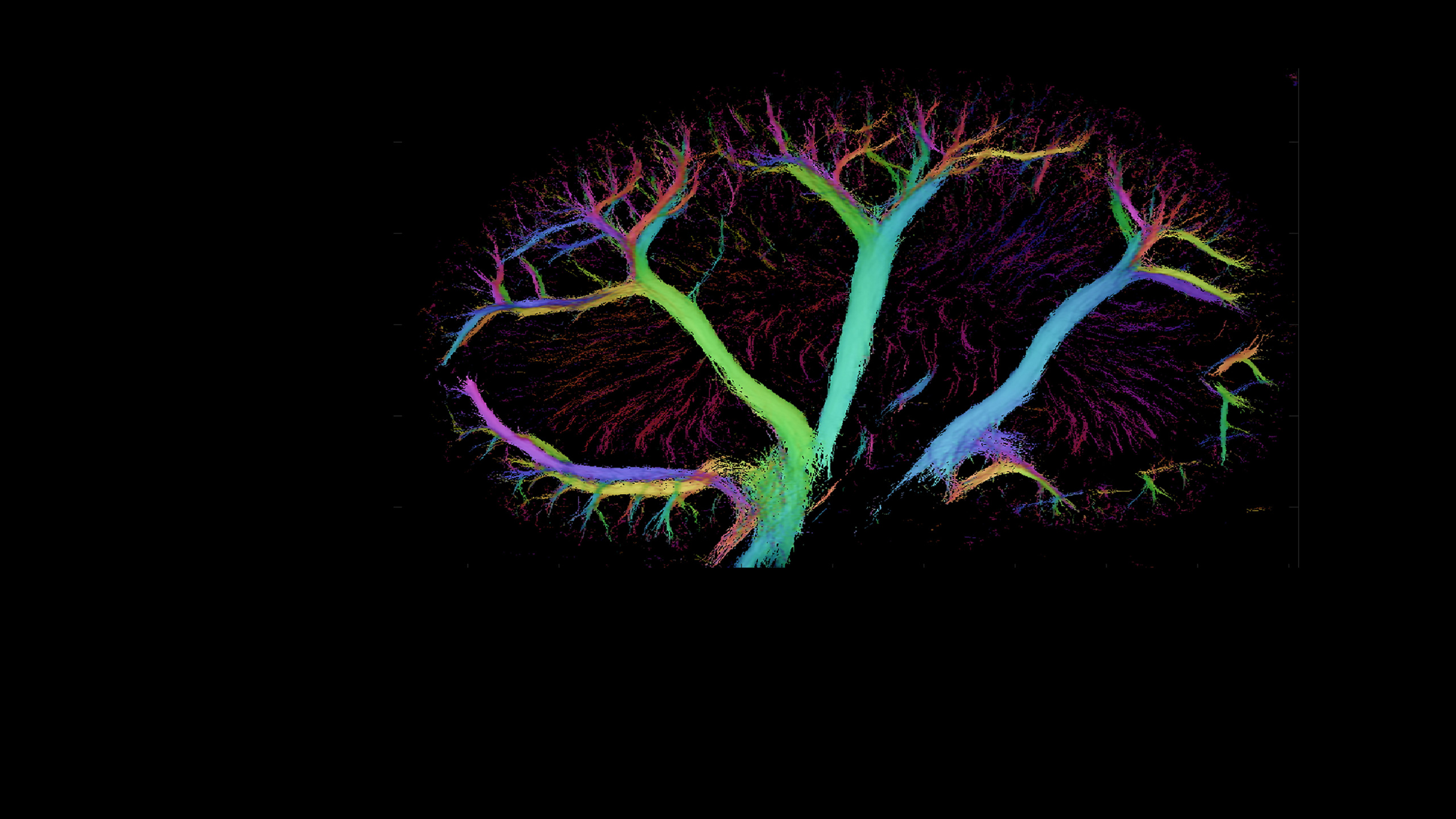
At CFU, we strive to develop new techniques, and improve existing ones, to yield more precise images of the complex flow patterns. This includes techniques for three-dimensional (3-D) vector flow imaging as illustrated. Another focus area is to make faster flow images and do real-time vector flow imaging to visualize the various dynamics in the human circulatory system.
This is pivotal as it is well established that flow patterns change rapidly throughout the cardiac cycle. The direction and magnitude also changes depending on the spatial location. However, conventional methods only estimate flow components towards or away from the transducer. This poses a huge challenge for quantitatively measuring the magnitude (and direction) of the blood’s velocity. This problem has been acknowledged for many years and a large number of techniques have been proposed to solve this.

Several years ago, The Transverse Oscillation (TO) method was developed at CFU - a method for estimating the velocity vector in the image plane. The method has been implemented by BK Medical on their commercial scanners and has been FDA approved for clinical use. Nowadays, the research focuses on expending the TO technique to other transducer geometries and improving the precision. Effort is also put into implementing the technique in OpenCL to allow real-time visualization using the experimental research scanner SARUS for data acquisition.
The TO approach has been extended to full 3-D imaging by using a 2-D matrix transducer. The technique has been implemented on SARUS and measurements have been conducted on a circulating flow-rig, on phantoms and in-vivo. This is still an active research area with focus on visualizing the complex flow patterns and proving quantitative measures of the volumetric flow rates in-vivo.
Effort is also being put into the developement of algorithms using synthetic aperture (SA) or synthetic aperture sequential beamforming (SASB) in combination with directional beamforming and duplex imaging techniques to yield high-frame rate flow imaging. The purpose is to expand the dynamic range of flow estimators to allow visualization of both rapid and slow flow.
Research within this area is performed by
Imaging

Ultrasound imaging is one of the most popular diagnostic methods - it is a non-invasive and low cost technique.
However, there are certain limitations. In conventional ultrasound, the recording of images is sequential in one direction at the time. This limits how many images can be shown per second, which is especially problematic when three dimensional imaging is desired, as in scans of the heart.
At CFU we are researching extensively in the ultrasound technique of the future - synthetic aperture – which should make it possible to obtain a better resolution, contrast and depth penetration.
Other research projects concentrate on methods to improve image quality by reducing speckle - the result of the constructive and destructive coherent summation of ultrasound echoes - and through optimizing operator-dependent controls.
Group Leader
Jørgen Arendt Jensen Head of Section, Professor, Ph.D., Dr.Techn. Department of Health Technology Phone: +45 45253924 Mobile: 40 42 51 50 jaje@dtu.dk


