Teaching in courses:
Research Group
MEMS Applied Sensors Group
Group leader: Erik Vilain Thomsen
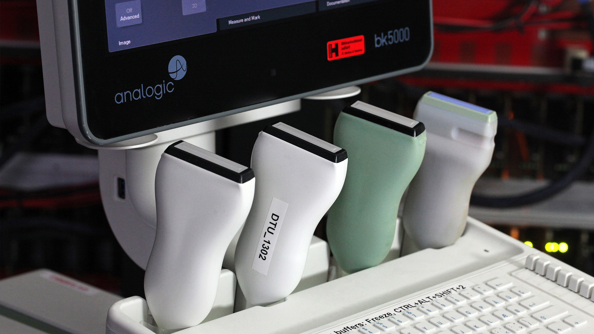
The scientific efforts of the MEMS group are focused on advancing medical ultrasound imaging by researching capacitive micromachined ultrasound transducers (CMUTs) as an alternative to the commonly used piezo electric transducers.
The MEMS group has developed both linear array and row-column based CMUT transducers fabricated using silicon based nano and microfabrication. Major accomplishments include:
- Development of robust and stable processes for CMUT fabrication in the DTU Nanolab cleanroom for both fusion and anodic bonded devices
- Development of analytical and finite elements models for design of CMUT arrays
- Fabrication of six generations of linear array medium frequency probes and one high frequency probe
- Development of five generations of row-column transducers for 3-D ultrasound imaging using integrated apodization for the first time
- Established a laboratory for characterization of CMUT devices both on wafer level and on single chip
- Established a packaging laboratory for assembly of ultrasound probes

Research vision
The UN defined Sustainable Development Goals work as a guiding star to achieve a better and more sustainable future for all. Working with MEMS and Healthcare Engineering the UN Goal 3 - "Ensure healthy lives and promote well-being for all at all ages" - guides us towards the future. We believe that the MEMS group and our collaborators can make an important contribution to meet this challenge by advancing the field of medical ultrasound imaging. Good health and well-being for all requires increased access to physicians all over the world and these physicians need technology for diagnosis and cure. Medical ultrasound is rather inexpensive compared to other image modalities such as MR and CT, and ultrasound systems are portable making them very well suited for use also at remote sites. Our research vision is to generate new ultrasound probes and systems for inexpensive 3D medical imaging together with other experts in the field. The vision is to be able to image in real time internal body structures and monitor flow at the capillary level in full 3D without using contrast agents. Such a system will enable advanced ultrasound imaging to be performed using inexpensive equipment and will help to provide advanced healthcare for all at a sustainable cost, i.e. support access to quality essential health-care services (UN target 3.8).
Research Projects
In the ”Super resolution Ultrasound Imaging (SRI)”, funded by the Innovation Fund Denmark, the MEMS group work with the company BK Medical and a range of collaborators, including Prof. Jørgen Arendt Jensen from DTU, on advancing 3D imaging using advanced row-column addressed CMUT probes to track the movement of injected contrast agents.
To realize SRI without using contrast agents is a challenging task that requires interdisciplinary research at the highest level. To fulfill this ambitious goal the MEMS group, work closely with a range of top experts in the field including
- Prof. Jørgen Arendt Jensen heading the Center for Fast Ultrasound (CFU) at the Technical University of Denmark (DTU) - he is an expert in medical ultrasound imaging
- Prof. Mikael Bachman, Department of Diagnostic Radiology, Ultrasound, Rigshospitalet -he is an expert in clinical ultrasound and cancer
- Associate Professor Charlotte Mehlin Sørensen, Department of Biomedical Science, University of Copenhagen - she is an expert in renal physiology and a specialist in performing experiments on living rodents.
Together we share a common vision for the next ten years and our research is funded by the ERC Synergy grant ”SURE: 3D Super resolution Ultrasound Real time imaging of Erythrocytes” where we research how to track the movement of the red blood cells (Erythrocytes).
SURE
The SURE project, "3D Super resolution Ultrasound Real time imaging of Erythrocytes", is funded by an ERC Synergy grant. The project will develop and research a new super resolution ultrasound imaging method capable of resolving 3D capillary flow in the human body.
The approach tracks the motion of the individual red blood cells (erythrocytes) in a three-dimensional volume for a full visualization of anatomy, flow, and perfusion in a volume down to approximately 13 cm at a rate of 20 volumes per second.
The SURE imaging approach aims to yield a paradigm shift in the scientific study, diagnoses, and treatment of cancer, diabetes, and vascular diseases at the capillary level, as it enables the possibility of volumetric visualizing capillary perfusion in real-time at frame rates above 20 Hz without injection of contrast agents. Imaging is performed using ultrasound at normal diagnostic levels with no known adverse effects and can, thus, be used on a wide range of the population from newborns to the elderly for both diagnosis and repeated screening.
The super resolution imaging is performed without using contrast agents and is thereby expected to be several thousand times faster than current methods. The method is expected to have an isotropic resolution of 50 micrometer in all directions, and the smallest details visible is thereby 100-400 times smaller volumetrically than current state-of-the-art 3D ultrasound imaging. Using deep learning is expected to further advance detection of targets making a resolution of 10 micrometer possible in flow measurements. These highly ambitious goals can only be attained in a synergistic research effort, and therefore the SURE project combines knowledge from several research groups.
The scientific project includes breakthroughs in silicon row-column probes with high element count, advanced synthetic aperture ultrafast coded imaging, deep learning for detecting and tracking of cells, pressure gradient estimation, and visualization and quantification of several hundreds of Gbytes volumetric data. The research finally leads to clinical trials conducted on rodents and humans for studying the changes in perfusion for diabetes and cancer and reveal the efficacy of SURE.
The role of the MEMS group in this project is to research and develop the advanced CMUT probes needed for the project. To achieve a resolution high enough to track individual erythrocytes it is estimated that these RCA probes will be required to have 1000 rows and 1000 columns and making such a transducer is a formidable task. However, based on long experience in working with MEMS and transferring MEMS technologies to production and to clinical research combined with the excellent facilities at DTU (e.g. the DTU Nanolab cleanroom, the SARUS research scanner at CFU, access to high performance computing, and the MEMS laboratory) we feel confident that it is realistic, although difficult, to achieve this ambitious goal.Super resolution imaging - SRI
Super resolution ultrasound imaging (SRI) is a new groundbreaking method enabling a true leap in diagnosis of cancer and diabetes. It can potentially attain a 10 micrometer resolution by using ultrasound tracking of contrast agents and opens a whole new and exciting field for
investigating human microvascular physiology and diagnostics. The project, supported by Innovation Fund Denamrk, translates SRI from experimental 2-D imaging of immobilized rodents, to human clinical use in full 3D.
The project partners develop new silicon based probes and 3D printed ultrasound phantoms (this research is done by the MEMS group), a new flexible scanner architecture, imaging and motion estimation schemes, and conducts human trials on cancer and diabetes patients. This makes it possible to introduce new, inexpensive 3D imaging schemes based on a new flexible software scanner and advanced silicon based probes on the market. These are used for SRI and 3D abdominal ultrasound imaging with a full 3D visualization of flow with the potential of radically improving vascular imaging, cancer staging and diabetes quantification.
The aim of the SRI project is to create a proof of concept prototype of a super resolution ultrasound imaging scanner able to detect, visualize, quantify, and assist in diagnosing the microvasculature of tissue and lesions, both malignant and benign in realistic models in a preclinical trial.
The role of the MEMS group in this ongoing project is research and development of RCA CMUT probes for use both in rodents and humans and the 3D printed flow phantoms needed for calibration.
Laparoscopy
Medical ultrasound imaging is widely used in the clinic for diagnostic purposes because it is an easy to use, safe and inexpensive technique as compared to CT and MR scanning.
The ultrasound imaging technique is, however, also used during surgery where specialized ultrasound probes have been developed for applications such as laparoscopy. Laparoscopy is a minimally invasive technique where surgery in the abdomen or pelvis is performed using a laparoscope with a built-in camera to guide the surgeon. Laparoscopic ultrasound transducers can be utilized to visualize hepatocellular lesions or liver metastases. During surgery, ultrasound is used to guide biopsies and ablation techniques, and assist in assessing the extent of benign and malignant lesions prior to laparoscopic resection or ablation. In the gall bladder, the main use of ultrasound is as a potentially faster and more cost-effective alternative to intraoperative cholangiography in identifying choledocholithiasis.
The adoption rate of ultrasound in surgery and intervention has been low due to difficulty in interpreting the 2D images generated by ultrasound and the lack of contrast in the images. Therefore, intraoperative CT and MRI have been gaining ground and are dominating the market. These multi-million dollar installations are characterized by high operational cost, slow imaging which inhibits the workflow but provides the much-needed 3D navigation. With the development of a real time 3D intraoperative ultrasound solution, the surgeon will be equipped with fast, cost efficient and accurate imaging system which can guide the surgery in a matter of minutes rather than the 30-60 minutes that the CT and MRI will require to generate the same 3D model of the organ.
The main goal of the project is to develop imaging using a Capacitive Micromachined Ultrasound Transducer (CMUT) based laparoscopic 3D ultrasound transducer with a coded excitation scheme optimized for Contrast Enhanced UltraSound imaging (CEUS). The news value includes
- Accurate 3D ultrasound model of organ for real-time navigated surgery (competing imaging modalities are currently based on competing CT and MR which takes around 30-60 min
- New coded excitation schemes for CMUT based imaging and CEUS
- First CMUT based 3D laparoscopic ultrasound transducer.
The project is a collaboration between DTU and the company bk medical supported by the Innovation Fund Denmark.
Group Leader
Erik Vilain Thomsen Professor Department of Health Technology Phone: +45 45255766 Mobile: +45 21363016 ervt@dtu.dk