Research Group

Scalable organ on a chip systems

Group Leader: Martin Dufva

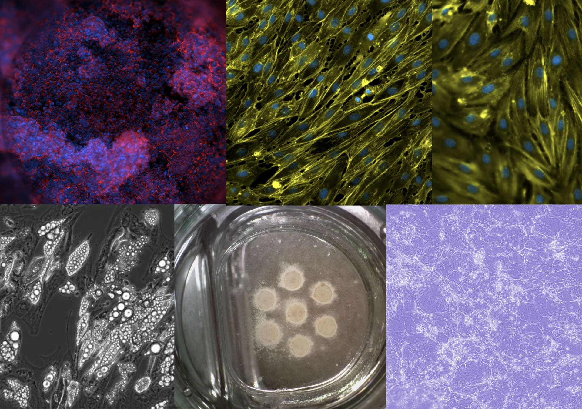
In the Scalable organ-on-a-chip system group, we aim to build advanced connected tissue models for in vitro testing of medicinal products. The application is drug development in the pharmaceutical industry and precision or personalized medicine at hospitals. However, any tests, such as toxicology, are possible applications. Our vision is to replace experimental animals with complex organ system with better predictive value. To achieve this, we apply minimal necessary engineering of organ-on-a-chip technology to obtain advanced cell culture systems that adhere to industry standards, automation, and instrumentation, and is furthermore mass producible. Through our collaborators, we are working with the intestine, liver, immune system, blood vessels, blood brain barrier, brain, fat, and cancer cells.

Some tissues are modeled using differentiation of induced pluripotent stem cells and mesenchymal stem cells while others are modelled using primary and immortalized cell lines. During prototyping, we apply 3D printing of hard materials that we subsequently modify with various hydrogels. The hydrogels are populated with cells on the inside or outside to mimic a tissue slice. Multiple of these can then be stacked on top of each other to model complex multiorgan systems, such as the gut – brain axis, for studying and interfering with virus transduction or the first pass metabolism. Shear and improved mass transfer is obtained using gravitational flow that is easily scalable. 
Research projects

Modular co-cultures of organ and tissue models

We develop interconnected organ models for biomedicine by stacking inserts on top of each other (Jepsen 2018, Dogan 2020). We are focusing on barriers and 3D tissues such as spheroids and densely packed sheets of cells modelling the intestine, blood vessels, liver and fat tissue. Through collaborations we also address many more organ systems. There is still much to learn about tissue and organ interactions and thereby how to recreate a representative in vitro organ system for replacing experimental animals.  

Techniques to design and fabricate automated cell culture systems

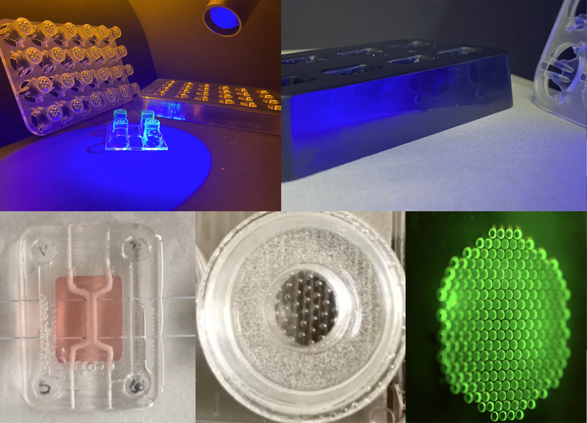
We design custom cell culture devices using 3D printing, or vacuum forming in polystyrene. 3D printing allows for much more complex devices than vacuum forming of polystyrene, but the latter material is traditionally used in cell culture plastics which is a clear advantage regarding biocompatibility and industrialization. We prototype vacuum forming molds using 3D printing and later we translate these to polished aluminum. Examples of custom inserts are 24-, 48-, 96-well plates as well as a range of devices that can be actuated using rockers to, for instance, align endothelium and activate epithelium.   

Understanding the cell physiology of in vitro models 

The devices we use are simple, but scalable, compared to many organ-on-a-chip devices and the mass transfer in these new devices is not well studied. Neither is the medium-usage pattern, nor the effects of endothelial or epithelial barriers on separating two mediums from each other. This becomes very important when using different mediums in stacked culture where two or more layers of tissue may be exposed to complex gradients derived from different mediums.