New unique techniques for investigating our intestinal immune system can lead to new possibilities for treatment of chronic intestinal diseases.
The intestinal immune system is essential for gut health, but when dysregulated it can lead to chronic diseases such as Crohn’s disease and ulcerative colitis.
The intestinal immune system can be divided into two functionally distinct immune compartments:
- the Gut-Associated Lymphoid Tissues (GALT), where adaptive immune cells are initially activated and differentiate into specific functional subsets;
- and the intestinal lamina propria, where activated immune cells localize and perform their effector and regulatory functions.
However, our current understanding of the immune composition and function of human GALT is limited, due to a lack of protocols to identify and isolate these compartments free from contaminating tissues.
New techniques
A research team headed by Professor William Agace, DTU Health Tech, have developed unique techniques to identify and isolate human GALT. Using these techniques, they described the distribution of GALT along the length of the human intestine and performed a detailed phenotypic and functional analysis of the adaptive immune cell compartment of human GALT as well GALT-free lamina propria.
“Our findings provide a first ‘roadmap’ of the GALT structure and composition along the length of the human intestine and additionally suggest that human GALT play an important role in driving region-specific adaptive immune responses within the intestine”, Professor William Agace says. And he continues, "This finding, which has previously been suggested from studies in mice, is particularly interesting as Crohn’s disease and ulcerative colitis affect distinct regions of the intestine, with Crohn’s disease primarily affecting the terminal ileum and proximal colon, and ulcerative colitis affecting the rectum and distal colon. We hypothesize that local adaptive immune activation in GALT may contribute to the highly regionalized inflammation observed in these diseases".
While GALT have been implicated in the development of both Crohn’s disease and ulcerative colitis, the role these structures play in the initiation and promotion of disease remains unclear.
Future studies using these novel isolation techniques will allow a direct comparison of the function of GALT in health and disease and may help elucidate novel disease-modifying pathways for the treatment of these chronic debilitating diseases.
The next step is to explore these aspects further in collaboration with Stanford University as part of the Gut Cell Atlas Consortium, based on a recent a grant from the Helmsley Trust, USA.
Read the full paper here.

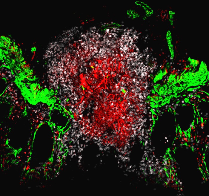
Image: Isolated lymphoid follicle of the human large intestine. T cells (white) and B cells (red) and an intestinal muscle layer called muscularis mucosa. The isolated lymphoid follicles extend through the muscularis mucosa muscle layer
Top image: Isolated lymphoid follicle of the human large intestine. Cells were stained in different colours, T cells (white), B cells (red), and germinal centers (green). T and B cells are two key immune cell types, germinal centers are important for the generation of antibody responses by B cells.