


Teaching in courses:
Research Group
Optofluidics
Group leader: Anders Kristensen
The integration and interplay between optical and fluidic functionalities defines the field of Optofluidics.
We do research in nano-optics and -fluidics for new sensing and actuation schemes. Our nanophotonics research is centered around optical metasurfaces and photonic crystals enabling ultra-thin, flat optical elements to address applications within biomedical devices. These optical elements are integrated with micro- and nanofluidics for compact sensor platforms. Our research is complemented by technology development within nanofabrication.
Projects
Ongoing research projects
Optical Metasurfaces
Nano-photonics and metasurface flat optics are directly related to the fundamental understanding of optics, established by Isaac Newton in the late 17th century, that light is carrier of information - frequency dependent amplitude (color), phase, polarization - which is coded into the electromagnetic field of light when it interacts with materials. Modern nanofabrication technologies are used to create artificial, man-made materials with engineered optical properties – offering new avenues to code information into light, without being constrained by the electromagnetic response of natural materials and their chemical compounds. In particular: Optical metasurfaces are two-dimensional arrays of nanoscale elements, called meta-atoms. The meta-atoms act as individual nanoscale optical antennas, which - via their size and shape – at the nano-scale offer full control of light as it interacts with the metasurface – for example when light is transmitted through the surface. In contrast to conventional technology, optical metasurfaces can provide a required, change to the optical field – amplitude, phase, polarization – abruptly, as it propagates over distances on the scale of the wavelength of light – less than a micron. This is used to create ultra-thin, so-called metasurface flat optics, including meta-lenses. In flat optics meta-lenses, the size, shape and morphology of the meta-atoms are varied across the surface area to obtain the variation of phase change required for the desired optical function.
We laser print metasurface flat optics by laser post-writing on template optical metasurfaces. Our optical metasurfaces are based on the concepts of both localized surface plasmon resonances (LSPR) and high-index dielectrics compatible with technologies for high volume manufactured plastic products. The optical metasurfaces are formed by nanoimprinting a surface texture comprising nanoscale cylinders or holes. By subsequent deposition of a thin film of metal or high index dielectric, isolated nano-disks or -cylinders are formed with designed optical resonances. Laser post-writing can modify nano-disks and holes, and hence the optical resonances. Laser pulses induce transient local heat generation that leads to crystallization, melting, and reshaping of the initial nanostructures. This enables flexible definition and alignment of optical components on high volume manufactured plastic products. Our approach offers a printing speed of 1 ns per pixel (in raster scan), resolution up to 127,000 dots per inch (DPI) and power consumption down to 0.3 nJ per pixel.
In the European Pathfinder project ODYSSEY, we exploit our on-resonance laser printing technology, enabling mass-customization of ultra-thin – so-called flat-optics – with a wide application field, for on demand manufacture of prescription eyeglass lenses.
Chromosome detection
Within the interdisciplinary synergy research project ChromoCapture we develop optofluidic technology capable of combined optical trapping and 3D imaging of metaphase chromosomes. The project is part of a collaboration with Prof. Ian Hickson and Prof. Eva Hoffmann at the Center for Chromosome Stability, Panum Institute, University of Copenhagen, and is funded under the NovoNordisk Foundation’s Interdisciplinary Synergy Programme.
We develop an automated multi-parameter analysis platform to investigate human chromosome abnormalities in real-time under an inverted microscope. The optofluidic platform combines three integrated sensing and actuation units (I) flow cytometry, (II) dual-beam optical tweezer, and (III) electrophoretic particle stretcher, combined with fluorescence imaging of the microscope.
The optofluidic platform comprises optical fibers aligned to a fused silica capillary microfluidic channel, assembled in a unit with external connections to microfluidic pump(s) and valves, fiber-coupled lasers and photodetectors and voltage source, and to be mounted on the microscope stage. The working principle is based on selective detection of the fluorescent light-emitting chromosomes with the flow cytometry unit before trapping at the optical tweezer region. An electric field is applied along the capillary channel for electrophoretic alignment and stretching of trapped chromosomes. Structural change of trapped and stretched chromosomes can be visualized by fluorescence microscopy.
Group Leader
Anders Kristensen Head of Sections, Professor Department of Health Technology Phone: +45 45256331 Mobile: +45 25171852 akri@dtu.dk
Teaching in courses
[Insert text here]
10031 Introduction to Engineering Physics
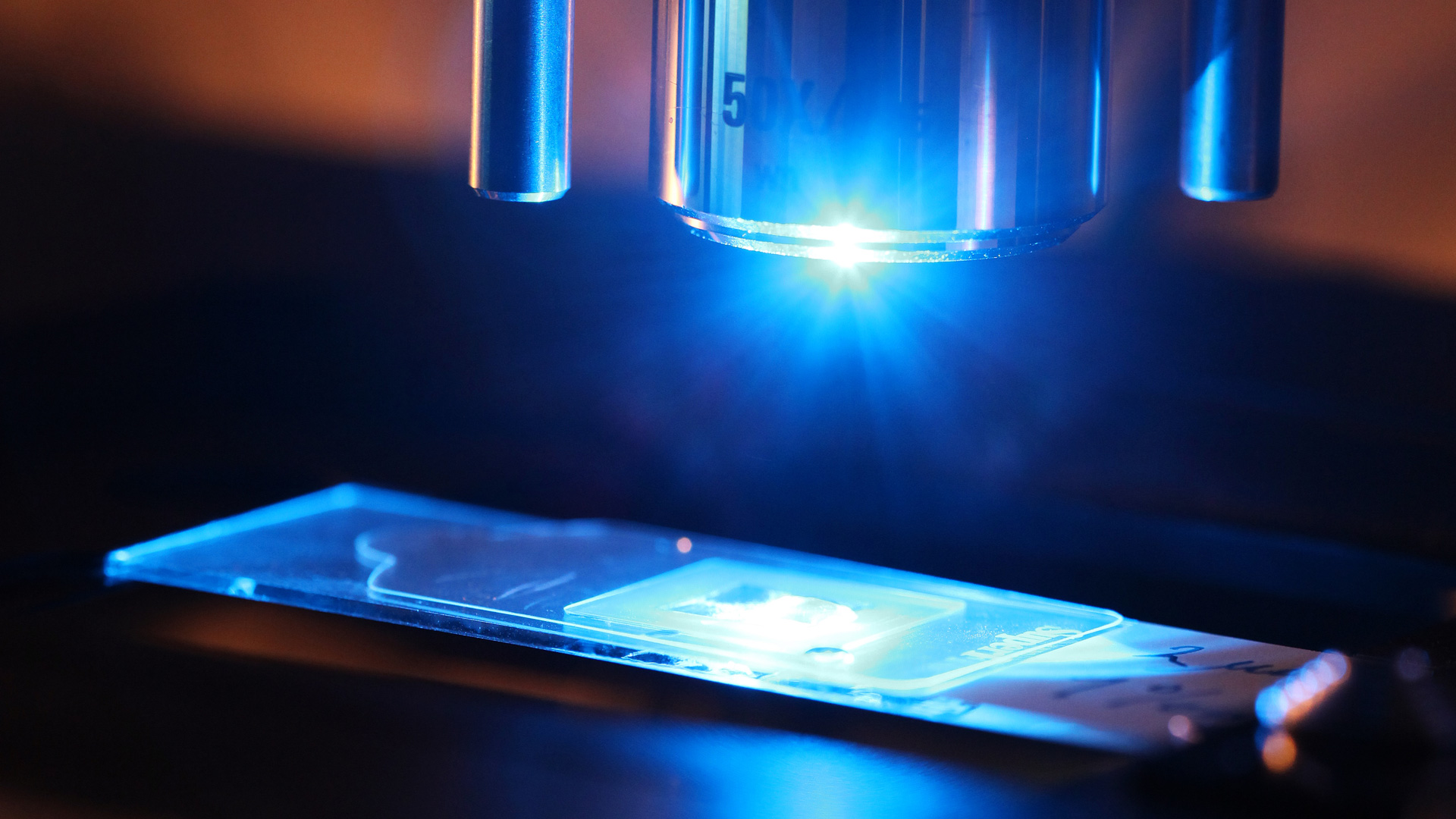
The lab

Equipment List
Light sources:
- two CryLaS FDSS532-150 diode pumped passively Q-switched solid state laser at 532 nm.
- CryLaS FTSS355-50 diode pumped passively Q-switched solid state laser at 355 nm.
- CrystaLaser DL405-25, CL532-025, CL660-025, 25 mW continuous wave lasers emitting at 405 nm, 532 nm, and 660 nm.
- Wicked laser Arctic, 1 W continuous wave GaN laser emitting at 445 nm.
- Ocean Optics HPX-2000 high power continuous wave Xenon light source (broadband 185 - 2200 nm).
- Newport, Oriel Instruments TLS-300X 300 W Xe Arc Lamp tunable light source (250 - 2400 nm).
- Thorlabs, Stabilized Tungsten-Halogen Light Source SLS201/M (300 - 2600 nm).
- Energetiq EQ-99XFC fiber coupled laser driven light source.
- High power LEDs M420L3 at 420 nm, M490L3 at 490 nm, M565L3 at 565 nm with drivers with pulse modulation DC2100.
- Cobolt Narrow Linewidth Laser 785nm, 500mW
- Toptica iBeam Smart 405nm, 150mW
Spectrometers:
- Acton SP-2756 Imaging Spectrograph with PIXIS100B Digital CCDcamera.
- Ocean Optics HR4000, model number HR4B208, wavelength range: 340 - 550 nm.
- Ocean Optics HR4000, model number HR4C1821, wavelength range: 550 - 620 nm.
- Ocean Optics HR2000, model number HR2A0328, wavelength range: 510 - 705 nm.
- Ocean Optics Jaz Spectrometer Module, wavelength range: 250 - 800 nm.
- Andor Shamrock 303, equipped with Newton 920 CCD
- Andor Shamrock 191, equipped with Andor IDUS CCD
High-resolution fluorescence microscope setup:
- Modified Nikon TE2000-U inverted fluorescence microscope with motorized prior stage and shutters. Light sources: 2 halogen, 1 metal-halide (fluorescence), 1 Hg (fluorescence backup). Filters: TRITC, FITC, 50/50 (UV-cut). The camera is a Photometrics Cascade II 512 back-illuminated electron multiplying CCD.
- A second Nikon Ti-U inverted fluorescence microscope. Light sources: 1 halogen, 1 metal-halide (epifluorescence), 1 Hg (transmition fluorescence). The camera is a Photometrics Evolve 512 back-illuminated electron multiplying CCD. DC power supply which can provide up to 150V (EA-PSI 6150-01 150 V/1.2 A) for electrophoresis. Houses a module that accepts fiber-coupled laser input sources for diffraction-limited focusing onto the sample.
- Both microscopes can be setup for confocal Raman spectroscopy.
Other equipment:
- Nano H50 series piezo electric stage (0.1 nm resolution, 50 µm travel, XY axes) suitable for mounting onto the group’s Nikon Ti series inverted microscope stages.
- 2 times 120 kHz DSP lock-in amplifiers SIGNAL RECOVERY model 7225.
- Hamamatsu X10468-04 SLM (Optical Phase Modulator).
- Tektronix TDS2014, 100 MHz 4-channel oscilloscope.
- Agilent 33220A arbitrary waveform generator.
- Thorlabs fast xy scanning stage MLS203-1.
- Sony microscope camera model DFW-SX910.
- Thorlabs high sensitivity USB 3.0 CMOS camera DCC3240C.
- Basler 60 fps camera acA1280-60gm.
- Harvard Pump 11 Plus syringe pumps, cat. number 70-2211.
- Two Fluigent MFCS-Flex pulseless pressure controllers, each with 8 independent channels, pressure range 1-1000 mbar, pressure resolution 1 mbar.
- One Fluigent MFCS-EZ pulseless pressure controller, with 4 independent channels, pressure range 1-350 mbar, pressure resolution 0.3 mbar.
Innovation
Patents filled:
A. Kristensen, K. Tølbøl Sørensen, Emil Højlund-Nielsen
"CUVETTE AND METHOD FOR MEASURING REFRACTIVE INDEX IN A SPECTROPHOTOMETER"
WO2017129196 (A1)
Peter Jan van der Zaag, Rodolphe Marie, Dianne van Strijp, Tom Olesen, Roland Vulders, Anders Kristensen
"MICROFLUIDIC DEVICE POSSESSING STRUCTURES ENABLING DIFFERENTIAL ANALYSIS OF A SINGLE CELL'S CONSTITUENTS"
WO2017085032 (A1)
A. Christiansen, A. Kristensen, N. Mortensen
"A NANOSTRUCTURED SURFACE FOR GREY SCALE COLOURING"
US2016202401 (A1)
J. Clausen, A. Kristensen, N. Mortensen, Emil Højlund-Nielsen, C. Jeppesen, A. Bruun Christiansen
"NANOSTRUCTURES FOR STRUCTURAL COLOURING"
US2016202394 (A1)
X. Zhu, A. Kristensen, N. Mortensen, Emil Højlund-Nielsen, C. Vannahme
"PHOTOTHERMAL MODIFICATION OF PLASMONIC STRUCTURES"
WO2016198657 (A1)
A. Kristensen, M. Rodolphe, J. Eriksen
"A Microfluidic Device with a Diffusion Barrier"
US2016137963 (A1)
A. Kristensen, M. Rodolphe, J. Eriksen
"A Microfluidic Device with Pillars"
US2016136642 (A1)
A. Kristensen, N. Mortensen, Emil Højlund-Nielsen, J Nørregaard
"AN OPTICAL DEVICE CAPABLE OF PROVIDING A STRUCTURAL COLOR, AND A CORRESPONDING METHOD OF MANUFACTURING SUCH A DEVICE"
US2016131808 (A1)
A. Kristensen, K. Mir, R. Marie, K. Rasmussen
"Microfluidic device and method for enzymatic processing of macromolecules"
DK2632592 (T3)
A. Kristensen, C. Vannahme, M. Dufva
"A SURFACE REFRACTIVE INDEX SCANNING SYSTEM AND METHOD"
EP3140636 (A1)
A. Kristensen, P. Hermannsson, C. Smith, C. Vannahme
"METHOD OF AND SYSTEM FOR IDENTIFICATION OR ESTIMATION OF A REFRACTIVE INDEX OF A LIQUID"
WO2015155349 (A1)
A. Kristensen, N. Mortensen, A. Christiansen
"A NANOSTRUCTURED SURFACE FOR GREY SCALE COLOURING"
DK201370657 (A)
K. Mir, R. Marie, A. Kristensen, K. Rasmussen
"Preparation and analysis of samples in micro-/nano-fluidic devices"
WO2012056192
J. Harries Hansen, H. O. Nielsen, A. Kristensen, M. B. Lindholm Mikkelsen, R. Marie
"Method for depositing sensor material on a substrate"
WO2011144652
M. Brøkner Christiansen, S. Ndoni, M. E. Vigild, A. Kristensen, P. Thomsen
"A nanoporous optical sensor element"
WO2011098090
R. Marie, A. Vig Laurberg, M. Brøkner Christiansen, A. Kristensen
"Solar energy harvesting system"
WO2011039356
T. Nielsen, B. Olsen, J. Nørregaard, A. Kristensen, K. Smistrup, E. Søgaard
"Injection molding tools with micro/nano-meter pattern"
WO2011038741
T. Nielsen, A. Kristensen, and O. Hansen
"A flexible nanoimprint stamp"
WO2006026993
S. Kragh, A. Menon, and A. Kristensen
“Optical amplification in miniaturized polymer cavity resonators”
WO2004112204
The company NIL Technology was founded by former students Theodor Nielsen and Brian Bilenberg in February 2006. The Company CPH Nano was founded by former students Kristian Tølbøl Sørensen and Emil Højlund-Nielsen